

📝 Author: Prof. Dr. Ömer Rıdvan Tarhan | 📅 Appointment: Isparta Meddem Hospital
Tru Cut Needle Biopsy
A tru-cut needle biopsy, also called core biopsy, takes cylindrical tissue samples from the lesion using a special needle. A tru-cut biopsy collects tissue, while a fine needle aspiration biopsy (FNAB) collects cells.
The tru-cut needle provides the pathologist with an undamaged tissue core from the lesion. To make an accurate diagnosis, the pathologist needs not only to examine the cells but also to assess their architectural arrangement within the tissue. In this respect, the tru-cut biopsy is superior to the fine needle aspiration biopsy (FNAB), which only collects cells. The lesion is typically a mass, and sometimes biopsies are taken from areas suspected on ultrasound. The procedure is performed using a specifically designed needle.
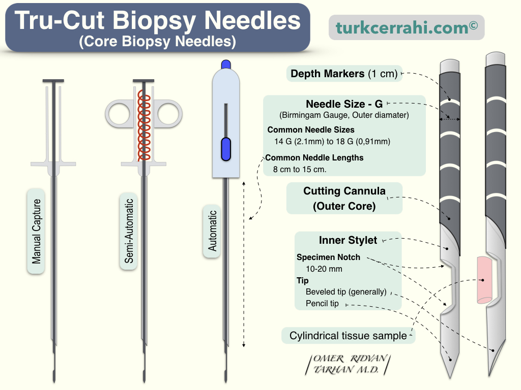
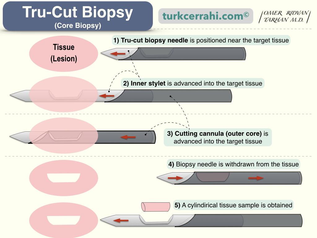
The principle is quite simple. A notched-edge needle is inserted into the lesion. Then, a sharp-edged metal sheath surrounding the needle is pushed toward its tip. The needle and sheath are attached to a spring mechanism. Depending on the device, the needle and sheath rapidly advance through the tissue in one or two presses. The sheath cuts the tissue corresponding to the hollow space in the needle, producing a cylindrical sample (core). Subsequently, the needle and sheath (cutting cannula) containing the sample are removed from the patient. This process is often repeated multiple times.
When dealing with palpable masses, a direct tru-cut biopsy can be performed. However, for especially non-palpable masses, the procedure can also be conducted under the guidance of ultrasound, computed tomography (CT), or magnetic resonance imaging (MRI).
How is Tru-Cut Needle Biopsy Performed?
Tru-Cut biopsy can be performed in the outpatient clinic under local anesthesia (ambulatory, outpatient, same-day procedure). A tru-cut biopsy is usually a very brief procedure, but when performed under imaging guidance (ultrasound, CT, mammography, MRI), it may take longer.
How is Tru-Cut Needle Biopsy Performed in the Breast?
If the biopsy is to be performed directly without the need for imaging, the patient lies on their back. If performed under imaging guidance, the patient may need to lie on their back, side, or stomach, depending on the imaging method used, or they may need to sit.
In all types of tru-cut biopsies, the skin area through which the biopsy needle will pass is numbed with local anesthetic. Then, a small incision (3-5 mm) is made in the breast skin using a pointed scalpel blade (no. 15) to create a passage for the needle. The tru-cut biopsy needle is inserted through this incision into the breast tissue to obtain a tissue sample. You may feel some pressure as the needle goes in. Imaging tests may also be performed to ensure the needle is directed to the correct location. After obtaining the tissue samples, the biopsy needle is removed. No stitches are needed. The area is covered with a sterile dressing. Applying pressure for a short period of time or using a compression dressing may help stop or prevent bleeding.
After Tru-Cut Biopsy: Limiting physical activities for about a day may be advised, but you can return to your usual activities after that. A true-cut biopsy may cause some bruising (ecchymosis), but it usually does not leave a scar. All biopsies can cause bleeding and swelling. The bleeding may cause the lump in the breast to appear larger after the biopsy. In most cases, this is not a cause for concern, and the bleeding, bruising, and swelling will fade over time.
Special Types of Tru-Cut Biopsies
Stereotactic Core Needle Biopsy
In this procedure, the doctor uses mammograms taken from different angles to determine the biopsy area. A computer analyzes the images and indicates the path the needle tip should take to reach the abnormal area. Stereotactic tru-cut biopsy is used to biopsy suspicious microcalcifications (tiny calcium deposits) or small tumors that are not clearly visible on ultrasound.
Vacuum-Assisted Core Biopsy
In a vacuum-assisted core biopsy, a hollow probe is inserted through a small incision into the abnormal area of breast tissue (guided by an imaging method). Then, vacuum (suction) is applied to the probe, and a rotating blade inside the probe cuts the tissues in a cylindrical (core) shape. This method allows for obtaining more tissue samples compared to a standard tru-cut needle.