

📝 Author: Prof. Dr. Ömer Rıdvan Tarhan | 📅 Appointment: Isparta Meddem Hospital
Where is the Abdominal Wall, What are its Layers?
The upper border of the abdominal wall is formed by the costal margins and the xiphoid process of the sternum; the lower border is formed by the upper margin of the pelvis (iliac and pubic bones); and the posterior border is formed by the vertebral column.
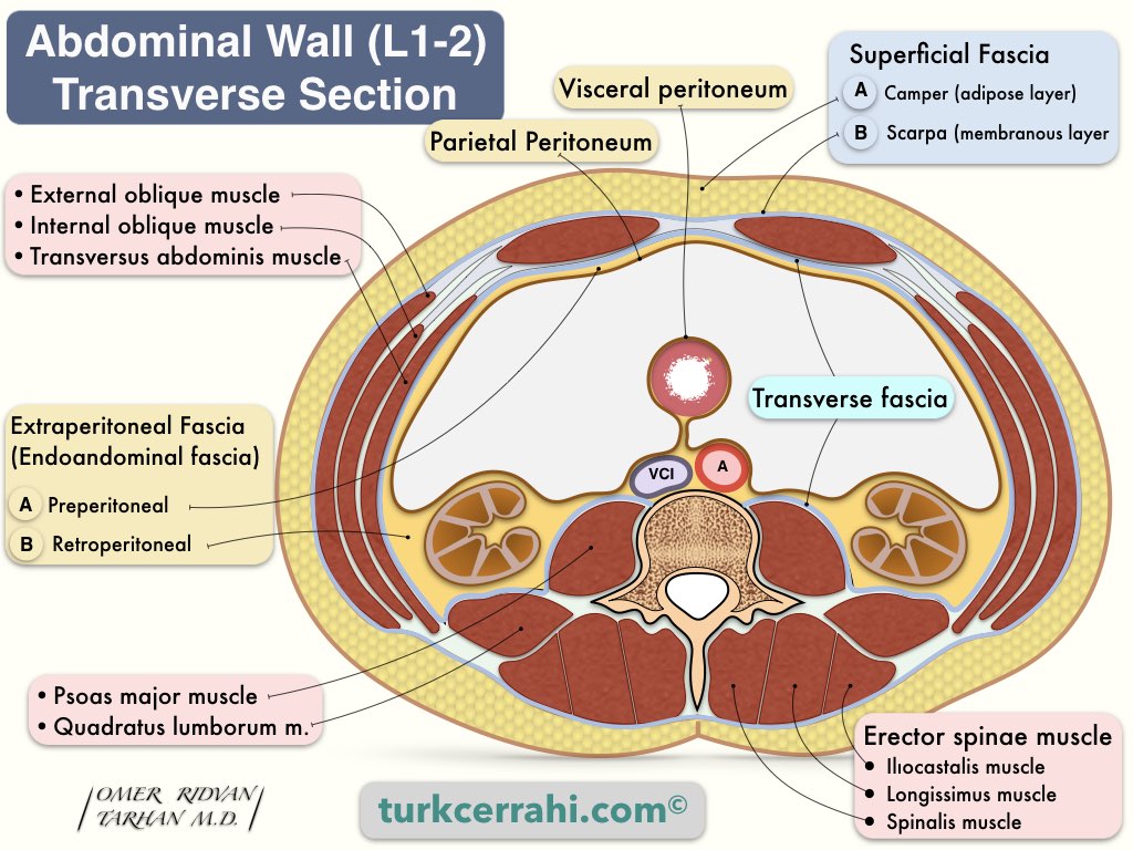
Layers of the abdominal wall (outside to inside)
- Skin
- Superficial fascia
- Superficial Layer (adipose, thick Camper's fascia)
- Deep Layer (thin, membranous Scarpa's fascia)
- Abdominal wall muscles and their aponeurosis
- Vertical Muscles: Rectus, Pyramidalis
- Flat Muscles: External oblique, internal oblique, transverse abdominal
- Fascia Transversalis
- Extraperitoneal Fascia (endoabdominal fascia)
- Peritoneum
Superficial Fascias of the Abdominal Wall (Camper’s and Scarpa’s Fascias)
Camper's Fascia: also known as subcutaneous adipose tissue. It is thinner in the upper abdomen and thicker in the lower abdomen. Camper’s fascia is the superficial layer of the superficial fascia.
Scarpa's Fascia: It is membranous and contains little or no fat. It starts below the umbilicus and continues into the thigh. Scarpa’s fascia is the deep layer of the superficial fascia.
Scarpa's fascia fuses with the fascia lata below the inguinal ligament. In males, it forms tunica dartos in the scrotum. Colles' fascia in the perineum is a continuation of Scarpa's fascia (superficial perineal fascia).
Scarpa's fascia is loosely attached to the aponeurosis of the external oblique muscle, while it is more densely attached to the linea alba and symphysis pubis, making it difficult to dissect during incisional hernia and abdominoplasty operations.
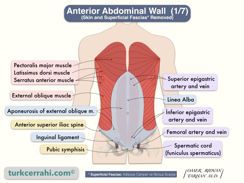
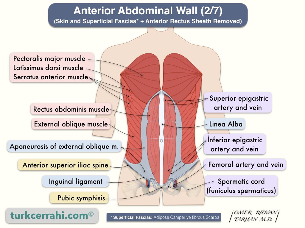
What are the Anterolateral Muscles of the Abdomen?
The anterior and lateral abdominal wall muscles are in five pairs.
- The two muscle pairs in the middle are vertical
- Rectus abdominis muscle
- Pyramidalis muscle
- Lateral muscles are flat and broad in 3 layers. From outside to inside;
- External oblique muscle
- Internal oblique muscle
- Transverse abdominal muscles
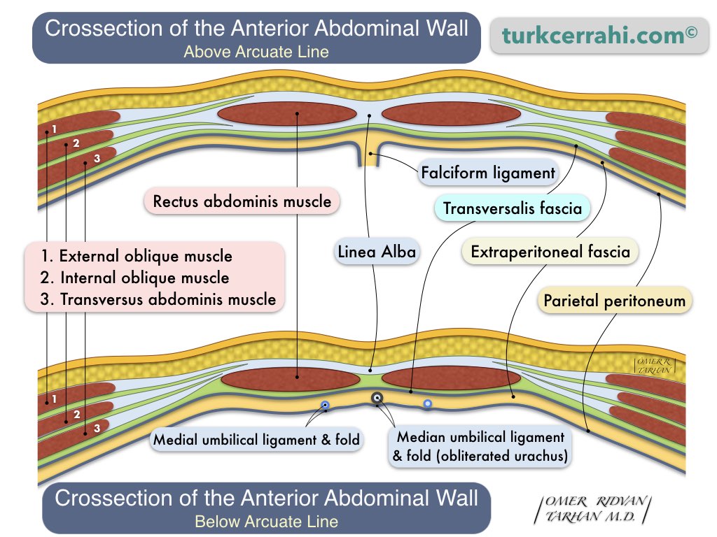
Although almost all of the abdominal wall is surrounded by muscles, there are no muscles in the midline (the linea alba, about 1 cm wide), and in the 4-5 cm section between the rectus abdominis muscle and the flat muscles, there are aponeuroses.
Functions of Abdominal Muscles: Movement of the trunk; keeping and protecting the abdominal organs (defense); coughing, vomiting, micturition (urination); defecation; birth.
Rectus Abdominis Muscle
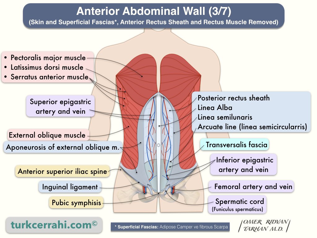
The rectus abdominis muscle begins broadly at the costal cartilages and the xiphoid process and narrows in the pubis symphysis. It is about 30 cm long and 5 cm wide. The rectus abdominis muscle is divided into 4-5 parts and interrupted in 3-4 places by fibrous bands (tendinous intersections). These are visible in well-developed muscles and weak people.
Pyramidalis Muscle
The pyramidalis muscles are about 4-5 cm long and form a narrow isosceles triangle. They are in front of the rectus muscle. Their base is attached to the anterior surface of the pubis and their apex to the linea alba.
External Oblique Muscle
The external oblique muscle is the most superficial of the three flat muscles. It is located just beneath the superficial fascia. Its fibers lie infero-medial. The external oblique muscle aponeurosis covers a large area of the anterior abdominal wall (in the middle). It passes in front of the rectus muscles and terminates at the linea alba.
Internal Oblique Muscle

The internal oblique muscle is in the middle of the external oblique and the transverse abdominal muscle. It is narrower than the external oblique muscle. Its aponeurosis bifurcates above the umbilicus, passes anterior and posterior to the rectus abdominis muscle, then fuses with the external oblique and transverse abdominal muscle aponeurosis, and terminates in the linea alba. Below the umbilicus, the internal oblique muscle aponeurosis passes in front of the rectus abdominis muscle as a single leaf, fuses with the aponeurosis of the external oblique muscle, and terminates in the linea alba.
Transverse Abdominal Muscle
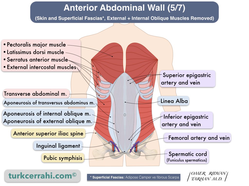
The transverse abdominal muscle is the innermost of the flat muscles. Above the umbilicus, its aponeurosis passes behind the rectus abdominis muscle, fuses with the posterior part of the internal oblique muscle aponeurosis, and terminates at the linea alba. Below the umbilicus, the transverse abdominal muscle aponeurosis passes in front of the rectus abdominis muscle, fuses with the aponeurosis of the internal oblique muscle, and terminates in the linea alba.
Extraperitoneal Fascia (Endoabdominal Fascia, Preperitoneal + Retroperitoneal Spaces)
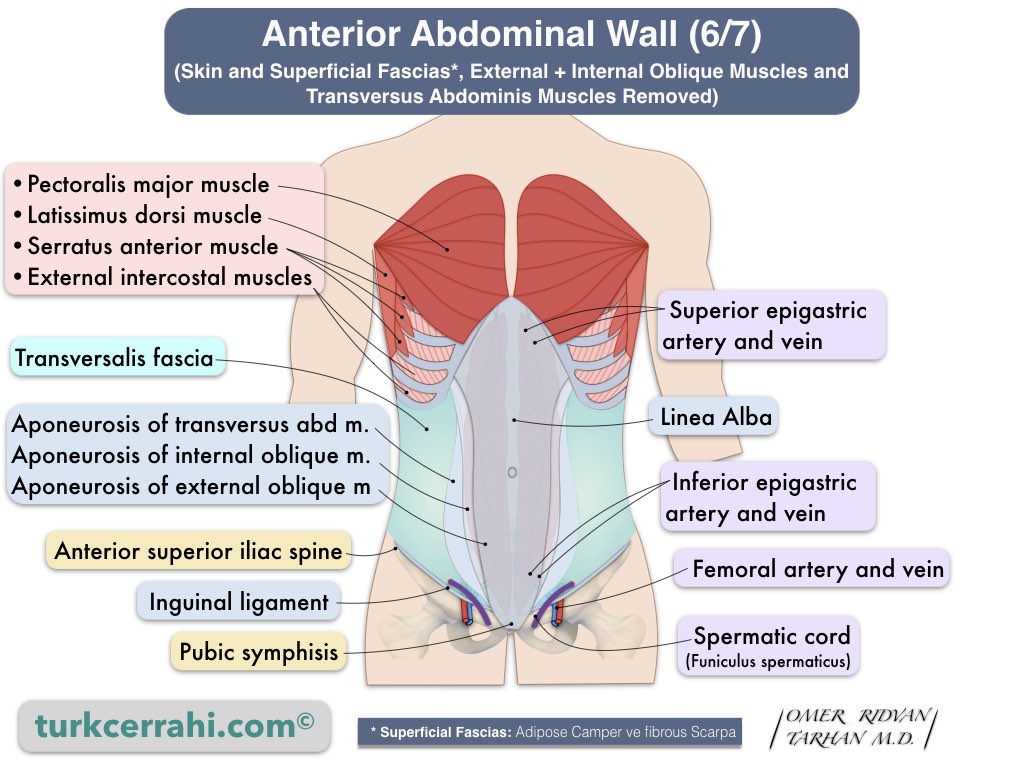
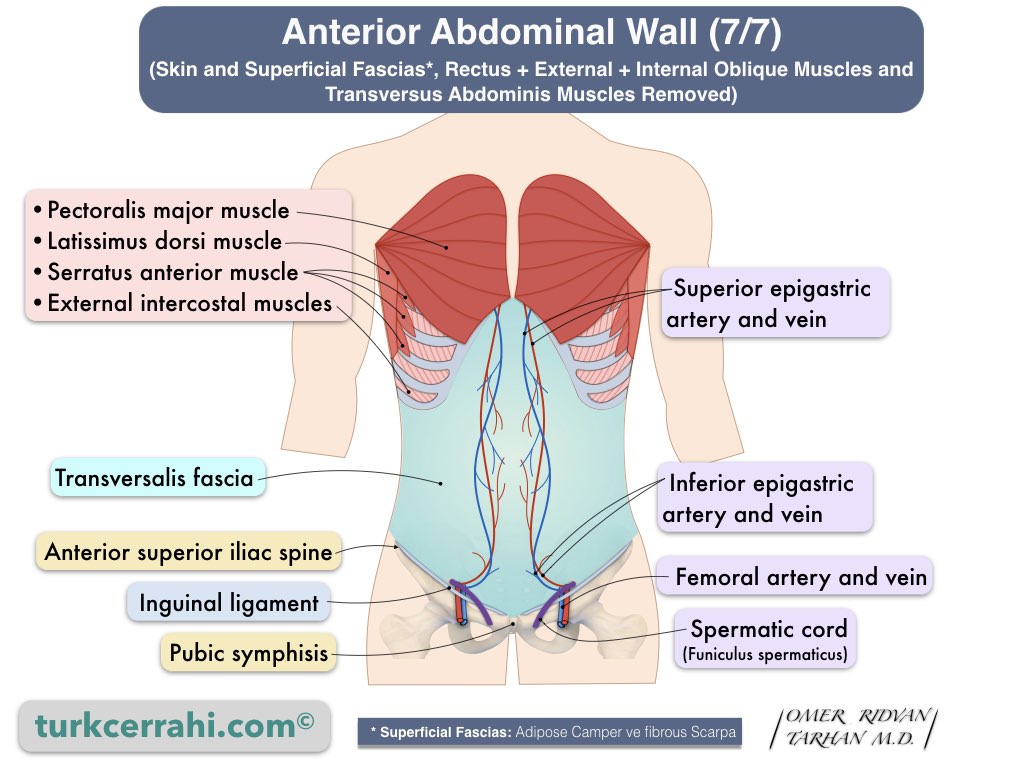
Extraperitoneal fascia is a connective tissue layer that is located between the peritoneum and the transverse fascia and contains abundant fat. Surgeons refer to the adjacent part of the anterior and lateral abdominal walls as the preperitoneal space, and the part on the posterior abdominal wall as the retroperitoneal space.
Peritoneum
The peritoneum is the innermost layer of the abdomen. It is a thin, serous layer lining the abdominal wall (cavity).
The peritoneum covers the organs completely (intraperitoneal organs) or partially (retroperitoneal and pelvic organs). The peritoneum that covers the abdominal cavity is called the parietal peritoneum, and the one that covers the organs is called the visceral peritoneum.
While the peritoneal cavity is completely closed to the external environment in men, it is connected to the external environment through the fallopian (uterine) tubes in women.
Intraperitoneal Organs
- Stomach
- Appendix
- Liver
- Transverse Colon
- Duodenum (1st part only)
- Small Intestines
- Pancreas (tail only)
- Rectum (upper ⅓ only)
- Spleen
Retroperitoneal Organs
- Adrenal (adrenal) Glands
- Aorta and Inferior Vena Cava
- Duodenum (all except the 1st continent)
- Pancreas (whole except tail)
- Ureters and Bladder
- Colon (ascending and descending)
- Kidneys
- Esophagus
- Rectum (lower ⅔ part)