

📝 Author: Prof. Dr. Ömer Rıdvan Tarhan | 📅 Appointment: Isparta Meddem Hospital
Biopsy
A biopsy is the extraction of tissue, cells, or fluid for microscopic examination by a pathologist. It is usually performed to determine whether a tumor is malignant (cancerous) or benign. Some infectious and inflammatory diseases also require biopsies to determine the severity and stage of the disease.
Different types of biopsies serve specific purposes and are used based on the location and nature of the suspected condition. The extracted samples are then sent to a laboratory for microscopic examination and analysis by a pathologist to provide a diagnosis and inform further treatment decisions.
Types of Biopsies
Open surgical biopsy
- Incisional biopsy: removal of a portion of the mass
- Excisional biopsy: removal of the entire mass. For example, a lumpectomy
Needle biopsy
- Fine-needle aspiration biopsy (FNAB): also called a percutaneous biopsy. It is performed using a syringe with a black needle (22G). From solid tissue, cells are extracted and examined by spreading them on a slide. It is most often used to diagnose thyroid nodules and occasionally breast masses. It is also frequently performed on the prostate.
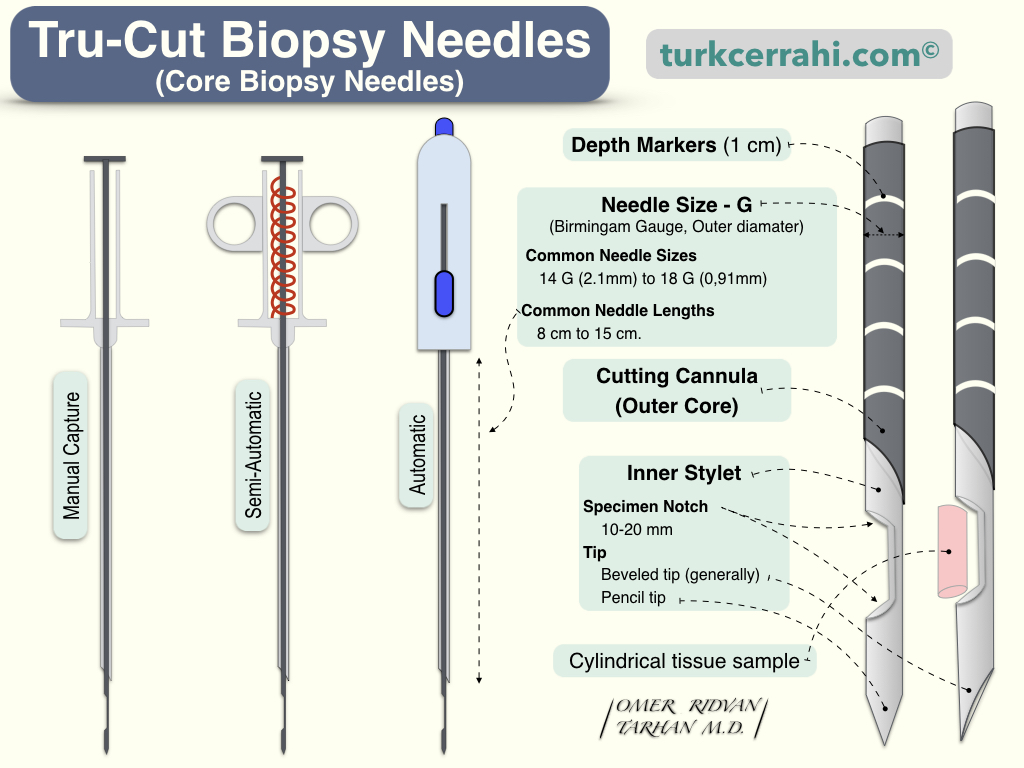
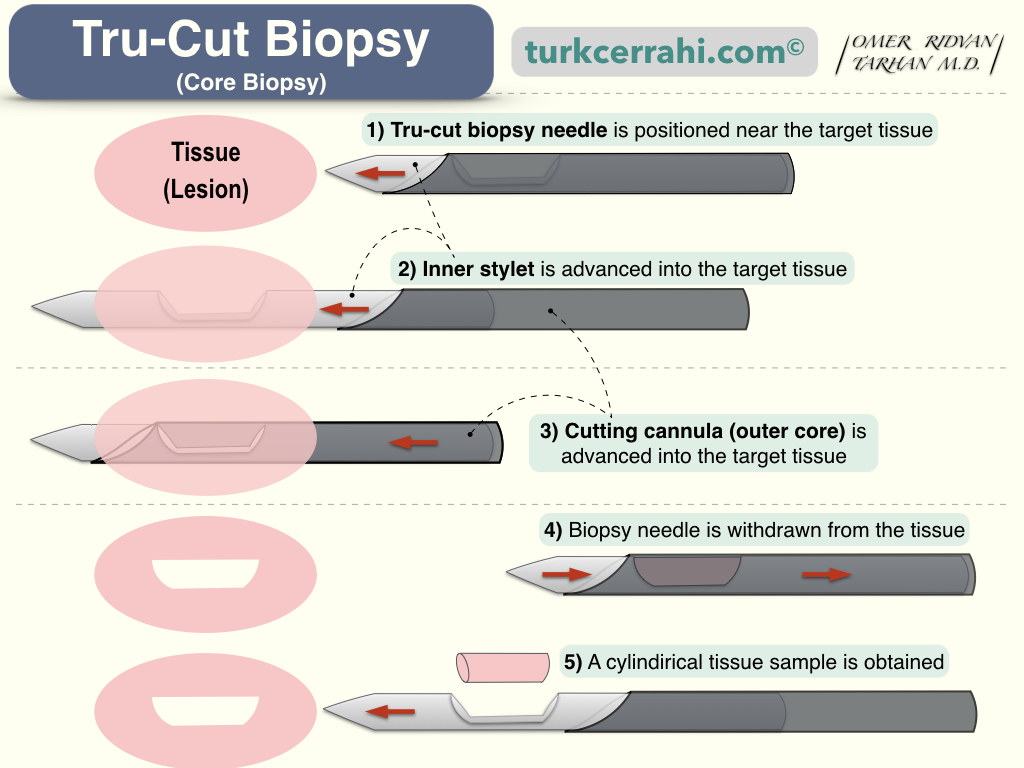
- Core needle biopsy: These needles have a mechanism that can remove cylindrical pieces of tissue from inside the mass. While FNAB obtains individual cells, a small piece of tissue is obtained with the core needle.
Brush biopsy
Using a small brush, a piece of tissue is scraped and then spread on a slide. Such as the bronchus and the renal pelvis. The brush is inserted through the working channel of the endoscope (bronchoscope) in order to perform a brush biopsy using an endoscope.
Chorionic villus biopsy
It is also known as chorionic villus sampling (CVS), and it is performed on pregnant women (10th to 12th week is optimal). This prenatal test can detect genetic and chromosomal abnormalities in the fetus. Chorionic villi are tiny, microscopic, finger-like projections that emanate from the placenta. The cells that make up the chorionic villi originate from the fetus; therefore, laboratory analysis can identify genetic, chromosomal, or biochemical fetal diseases. Chorionic villus sampling should be performed between the 10th and 12th weeks of pregnancy. The procedure can be performed transvaginally – transcervically, or transabdominally.
Cone biopsy
A cone-shaped piece of tissue is excised (removed) from the cervix of the uterus.
Endoscopic biopsy
It is performed with instruments passed through an endoscope’s working channel. Usually, small pieces of tissue are excised (punch biopsy) or polyps and similar structures are cut off from the root and burned (cauterized) using an instrument called a snare, and then they are grasped and removed with another instrument.
Sentinel lymph node biopsy
It is a procedure in which the lymph node with the highest likelihood of cancer cell spread in the tumor is identified, marked, removed, and sent for pathology analysis. The blue dye (isosulfan blue or Lymphazurin® 1%) or a radioactive substance is injected into the tumor, surrounding tissue, or the area of the nipple to mark the sentinel lymph node. The dye is transported by lymphatic vessels to the lymph node that is most likely to be metastasized by cancer first. The lymph node is then located using a gamma probe that detects radioactivity or a change in color (blue). If both techniques are employed, an incision is made at the point of greatest radioactivity, and the blue-stained lymph node can be easily extracted.
Shave biopsy
Shave biopsy entails removing the skin lesion as well as the top layer of skin by cutting parallel to the skin surface.
Stereotactic biopsy
It is a technique that uses a stereotactic system to determine the location of the biopsy area. It is typically utilized in the breast and the brain. A cube-shaped frame is screwed onto a patient’s head during a stereotactic brain biopsy. To determine the coordinates of the biopsy area relative to the frame, computed tomography is taken. A movable arc on the frame is directed to the target, and a needle is inserted through the arc to obtain a biopsy from the area. A stereotactic breast biopsy is also referred to as a wire localization biopsy.
Wire localization biopsy
A wire localization biopsy (stereotactic biopsy) is used for lesions in the breast that are non palpable but visible on a mammogram or ultrasound. A fishing-hook-shaped wire is inserted into the lesion under ultrasound or mammographic guidance (the needle does not retract). The surgeon removes the tissue around the wire in a cylindrical shape under local or general anesthesia. Before the operation is finished, the removed breast tissue is examined with ultrasound or mammography to ensure that the lesion has been removed.
Sternal biopsy
A bone marrow biopsy from the sternum is obtained by puncturing it with a special needle (the tissue is drawn into the syringe by negative pressure).
Liver Biopsy
It can be performed percutaneously (via ultrasound or computed tomography), with a thick needle (tru-cut), or during surgery (wedge biopsy) from the liver’s edge.
Percutaneous renal biopsy (kidney biopsy)
After locating the lesion using ultrasound, computed tomography, or angiography, a needle is inserted through the skin to obtain a sample of kidney tissue. This method is also used to diagnose kidney failure, determine the prognosis of kidney disease, assess the extent of kidney damage, and determine the most suitable treatment. Urinary bleeding is the most common complication, which gradually subsides and disappears within a few days.
Percutaneous transthoracic needle aspiration biopsy
It is performed to obtain tissue samples from lung malignancies or other suspicious lesions. A radiographically-guided aspiration needle (e.g., a CT scan) is utilized. This procedure is generally contraindicated in patients receiving mechanical ventilation due to the risk of pneumothorax.
Punch biopsy
It involves removing a small piece of tissue (often from the skin) using a small, round cutting instrument (such as a punch or biopsy punch).
Vacuum-assisted biopsy
The procedure entails inserting a needle with a sharp-edged hole into the lesion. Negative pressure is applied to the needle, which is then moved back and forth to remove tissue fragments with sharp edges. These vacuumed tissues are placed in a container and sent to a lab for microscopic examination.
« Return to Dictionary