

📝 Author: Prof. Dr. Ömer Rıdvan Tarhan | 📅 Appointment: Isparta Meddem Hospital
Duodenum
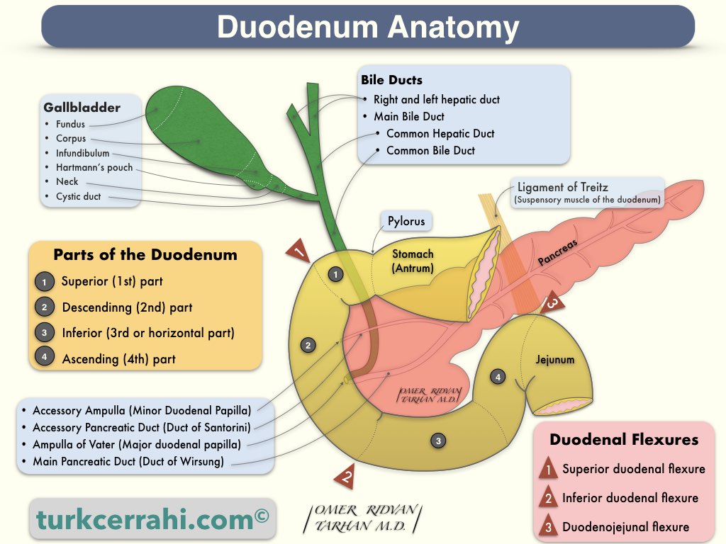
The duodenum is the first and widest section of the small intestine, which connects to the stomach at the pyloric sphincter and extends for about 25-30 cm (10-12 inches). It is a C-shaped tube that curves around the head of the pancreas and receives secretions from the liver, gallbladder, and pancreas through the hepatopancreatic duct. The bile duct and pancreatic duct do join together to form the hepatopancreatic ampulla (also known as the ampulla of Vater), which opens into the second part of the duodenum.
The primary function of the duodenum is to receive partially digested food from the stomach, neutralize the acidic contents with bicarbonate secretions from the pancreas, and further break down the food with digestive enzymes from the pancreas and bile from the liver and gallbladder.
Iron absorption primarily occurs in the duodenum. Duodenal ulcers are a common type of peptic ulcer. The duodenum is almost entirely retroperitoneal and is fixed in its location, unlike other parts of the small intestine.
Parts of Duodenum
The duodenum is anatomically divided into four parts, or sections, which are:
- The superior (first) part
- The descending (second) part
- The horizontal (third) part
The ascending (fourth) part:
1. The superior (first) part
This section is the widest and shortest part of the duodenum, measuring about 5 cm in length and attached to the liver by the hepatoduodenal ligament of the lesser omentum. The first 2 cm of this section is slightly dilated, forming the ampulla of the duodenum.The superior duodenal flexure marks the end of the first part of the duodenum.
2. The descending (second) part
This section is about 7.5 cm long and ends at the inferior duodenal flexure. The main pancreatic duct (Wirsung) and common bile duct open into the second part of the duodenum through the major duodenal papilla, or liver bud. This section also contains the minor duodenal papilla, where the accessory pancreatic duct opens.
The embryological border between the foregut and midgut is located just below the major duodenal papilla, although it is commonly and incorrectly believed to be at the level of the ligament of Treitz.
3. The inferior (horizontal, third) part
This section is about 10-12 cm long and runs transversely from the right side of the L3 vertebra to the left side of the L2 vertebra.
It is located anterior to the inferior vena cava, abdominal aorta, and vertebral column. The superior mesenteric artery and vein pass in front of this part of the duodenum. The duodenum can be compressed between the aorta and the superior mesenteric artery, leading to a condition called Superior Mesenteric Artery Syndrome, also known as Wilkie’s Syndrome.
4. The ascending (fourth) part
This section is about 2.5 cm long and ascends from the level of the L2 vertebra to the duodenojejunal flexure, where it becomes continuous with the jejunum.
The suspensory ligament of the duodenum, also known as the ligament of Treitz, is a fibromuscular band that suspends the duodenojejunal junction from the posterior abdominal wall. It is named after the Czech physician Václav Treitz, who first described it in 1855.
« Return to Dictionary