

📝 Author: Prof. Dr. Ömer Rıdvan Tarhan | 📅 Appointment: Isparta Meddem Hospital
Ascites
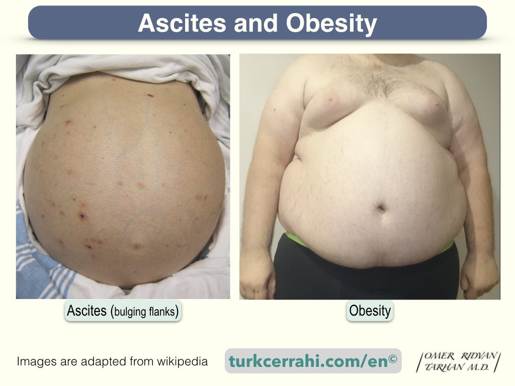
Ascites is the accumulation of fluid in the peritoneal (abdominal) cavity. Ascites is most commonly associated with liver disease, such as chronic liver disease (cirrhosis). Heart and kidney failure, peritonitis carcinomatosa, ovarian cancer, and metastatic liver cancer are other common causes. Technically, any fluid accumulation in the peritoneal cavity greater than 25 ml is considered ascites.
- Ascites Image retrieved from Wikipedia on 05 June 2023 under Creative Commons Attribution-ShareAlike 3.0 Unported. By James Heilman, MD – Own work, Image’s Page
- Obesity Image retrieved from Wikipedia on 05 June 2023. FatM1ke the copyright holder of this work, release this work into the public domain. Image’s Page
Diagnosis
Ascites is typically diagnosed through a physical examination, imaging tests such as an ultrasound or CT scan, and analysis of the fluid obtained through a procedure called paracentesis.
Physical Examination
- Abdominal distension
- Bulging flanks
- Shifting dullness
- Fluid wave test
Abdominal distension: The abdomen appears swollen and feels tense or tight due to the accumulation of fluid.
Bulging flanks: When the patient lies on their back, bulging of the flanks may be observed due to the accumulation of fluid in the dependent areas of the abdomen.
Shifting dullness: By tapping or percussing the abdomen, the healthcare provider can identify a change in the sound produced when the patient changes position. In ascites, there is a shift in the area of dullness (where the sound is muffled) towards the dependent, fluid-filled parts of the abdomen.
Fluid wave test: The healthcare provider will place their hand on one side of the abdomen and tap the other side. If a fluid wave is felt or observed, it indicates the presence of fluid in the abdomen.
Types of Ascites
| Transudative Ascites | Exudative Ascites | |
|---|---|---|
| Protein Levels | Low (less than 2.5 g/dL) | High (greater than 2.5 g/dL) |
| LDH Levels | Low | High |
| Cell Count | Low | High, primarily white blood cells |
| Specific Gravity | Low | High |
| Inflammatory Markers | Low | Presence of inflammatory markers |
| Etiology | Increased hydrostatic pressure or fluid homeostasis imbalances | Inflammation, infection, or malignancy |
Effusions can be classified based on their fluid composition as transudative or exudative ascites. Ascites is also a type of effusion and is also known as “peritoneal effusion”.
Light’s Criteria
Light’s Criteria is a set of three criteria used to differentiate between transudative and exudative pleural effusions. The three criteria of Light’s criteria are as follows:
- Pleural fluid protein to serum protein ratio (pleural protein/serum protein): A ratio greater than 0.5 is considered exudate.
- Pleural fluid LDH to serum LDH ratio (pleural LDH/serum LDH): A ratio greater than 0.6 is considered an exudate.
- Pleural fluid LDH level: If the pleural fluid LDH level is greater than two-thirds of the upper limit of normal for serum LDH, it is considered an exudate.
If any one of these criteria is met, the pleural effusion is classified as an exudate. If none of the criteria are met, the effusion is classified as a transudate.
Serum to Ascites Albumin Gradient (SAAG)
It’s important to note that Light’s criteria are primarily applicable to pleural effusions and may not directly apply to other types of effusions, such as peritoneal effusions (ascites).
The serum-to-ascites albumin gradient (SAAG) is a more accurate and physiologically relevant test for differentiating the causes of ascites. The SAAG is calculated by subtracting the peritoneal fluid albumin level from the serum albumin level.
Transudative ascites (SAAG <11 g/L)
Transudative ascites occur when the fluid accumulation is primarily due to increased pressure in the blood vessels or a disturbance in the fluid balance. It is commonly caused by liver cirrhosis, heart failure, and kidney disease and is characterized by a low protein content.
- Liver cirrhosis; responsible for 80% of cases of ascites,
- Heart failure; 3%,
- Budd-Chiari syndrome
Exudative ascites (SAAG >11 g/L)
Exudative ascites result from inflammation or infection in the peritoneal cavity, leading to the leakage of high-protein fluid. It is commonly associated with conditions such as peritonitis, pancreatitis, malignancies, or tuberculosis
- Pancreatic ascites develop when the pancreatic duct (wirsung) is damaged in pancreatitis, pancreatic injuries, or when a pancreatic pseudocyst ruptures. Pancreatic fluid accumulates in the abdomen.
- Chylous ascites have a milky appearance due to lymphatic fluid leaking into the abdominal cavity. It develops due to trauma, abdominal surgery, or tuberculosis.
- Malign Ascites; accounts for 10% of all cases of ascites. It is often a result of peritoneal carcinomatosis or cancer that has spread (metastasized) from another part of the body (carcinomatosis peritonitis).
- Renal ascites (nephrotic syndrome); caused by low levels of albumin in the blood. Albumin is the most important protein in the blood plasma, and it functions to keep fluid inside blood vessels.
Types of Ascites (Based on Underlying Disease)
- Cirrhotic Ascites are the most common type of ascites and are associated with liver cirrhosis, which is often caused by chronic alcoholism, hepatitis B or C infection, fatty liver disease, or other liver conditions. In cirrhotic ascites, the liver’s ability to function properly is compromised, leading to fluid accumulation in the abdomen.
- Malignant Ascites are caused by cancers that spread to the abdominal cavity, such as ovarian cancer, gastrointestinal cancers, or cancers affecting the peritoneal lining. Malignant ascites may also occur due to the liver metastases.
- Tuberculous Ascites occur when tuberculous infection involves the peritoneum (peritoneal tuberculosis).
- Pancreatic Ascites is characterized by the leakage of pancreatic enzymes into the abdominal cavity, leading to inflammation and fluid accumulation. It is often associated with pancreatitis, pancreatic pseudocysts, or pancreatic duct disruption.
- Cardiac Ascites occur when there is an impairment in heart function, leading to elevated pressure in the veins that drain blood from the liver. This increased pressure causes fluid to leak into the peritoneal cavity.
- Nephrogenic Ascites is associated with severe kidney dysfunction or failure. Impaired kidney function leads to fluid retention and electrolyte imbalances, resulting in ascites.
Severity of Ascites
- Grade 1 or mild ascites: Mild, only visible on ultrasound and CT scans,
- Grade 2 or moderate ascites: Detectable on physical examination. A fullness and shifting dullness can be felt in the lumbar region,
- Grade 3 or severe ascites: Visible ascites, confirmed by physical examination.